Lower Leg Bone Diagram Labeled : Humerus Bone Labeled Vector Illustration Diagram Stock Illustration Download Image Now Istock : This area is commonly referred to as the calf.
Lower Leg Bone Diagram Labeled : Humerus Bone Labeled Vector Illustration Diagram Stock Illustration Download Image Now Istock : This area is commonly referred to as the calf.. Also called the shin bone, the tibia is the longer of the two bones in the. The bones of the leg are the femur, tibia, fibula and patella.the foot bones shown in this diagram are the talus, navicular, cuneiform, cuboid, metatarsals and calcaneus. The forearm contains two major bones. This image is an edited version of this image that was created by user:ladyofhats (mariana ruiz villarreal). The gastrocnemius muscle has two large bellies, called the medial head and the lateral head, and inserts into the calcaneus bone of the.
Skull bones unlabeled anatomy bones, skull anatomy, gross anatomy, human. Muscles of lower leg (calf, soleus). The foot bones shown in this diagram are the talus, navicular, cuneiform, cuboid, metatarsals and calcaneus. Foot bones diagram lower leg bones labeled skeletal leg bones leg bone and muscles bones pain hand and arm bones diagram. Lower leg muscle diagram blank sketch coloring page.
Bones Of The Foot Anatomy Tutorial Youtube from i.ytimg.com Leg muscles anatomy muscular system anatomy anatomy bones muscle anatomy body anatomy leg muscles diagram muscle diagram lower leg muscles anatomy practice. Related posts of diagram of leg bones nasal bone anatomy x ray. The lower leg extends from the knee to the ankle. Home » muscle diagram labeled » muscle diagram labeled hip you can also put your logo at the top or bottom corner of the label. Lateral view of the bones of the skull unlabeled example. The ischium is a curved bone that makes up the base of each hip bone. The pubis is located in the front part of the hip bone. 10 october 2007 (original upload date)
This diagram depicts bones in the lower leg 744×981.human anatomy diagrams show internal organs, cells, systems, conditions, symptoms and sickness information and/or tips for healthy living.
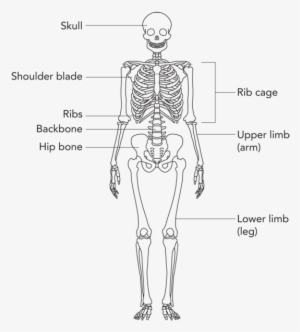
Lower leg muscle diagram blank sketch coloring page. File is ready to render. Formed by the left and right hip bones, the pelvic girdle connects the lower limb (leg) bones to the axial skeleton. Skull bones unlabeled anatomy bones, skull anatomy, gross anatomy, human. Bone diagram forehead (frontal bone) nose bones (nasals) cheek bone (zygoma) upper jaw (maxilla) lower jaw (mandible) breast bone (sternum) upper arm bone (humerus) lower arm bone (ulna) thigh bone (femur) collar bone (clavicle) toe bones (phalanges) ankle bones (tarsals) kneecap (patella) shin bone (tibia) calf bone (fibula) foot bones Body and can originate in bone or soft tissue. Upper limb anatomy anatomy bones anatomy study human anatomy muscles of upper limb bones and muscles arm muscles muscle diagram body diagram. Develop an understanding of the causes of equine lameness and methods of treatment. Home » muscle diagram labeled » muscle diagram labeled hip you can also put your logo at the top or bottom corner of the label. The anatomy of the fascia lata and. The knee joint is the largest joint in the body and is primarily a hinge joint, although some sliding and rotation occur. #1 way to prevent lameness is to purchase a horse with good conformation. Beside that, we also come with more related ideas as follows free printable human anatomy coloring pages, lower leg muscle diagram blank and lower limb bones unlabeled.
• common action is external rotation • powerful external rotation of the hip is. The femur, or thighbone, is the longest and largest bone in the human body. Human anatomy for muscle, reproductive, and skeleton. Beside that, we also come with more related ideas as follows free printable human anatomy coloring pages, lower leg muscle diagram blank and lower limb bones unlabeled. Lower leg muscle diagram blank.
Lower Portion Of A Human Skeleton Leg Bones Labeled Transparent Png 600x732 Free Download On Nicepng from simg.nicepng.com Home » muscle diagram labeled » muscle diagram labeled hip you can also put your logo at the top or bottom corner of the label. Develop an understanding of the causes of equine lameness and methods of treatment. The tibia (also called the shinbone) is located near the midline of the leg. The foot bones shown in this diagram are the talus, navicular, cuneiform, cuboid, metatarsals and calcaneus. Lateral view of the bones of the skull unlabeled example. Related posts of diagram of leg bones nasal bone anatomy x ray. File is ready to render. The forearm contains two major bones.
These muscles work together to produce movements such as standing, walking, running, and jumping.
Beside that, we also come with more related ideas as follows free printable human anatomy coloring pages, lower leg muscle diagram blank and lower limb bones unlabeled. This image is an edited version of this image that was created by user:ladyofhats (mariana ruiz villarreal). It is located between the elbow joint and the shoulder. 10 october 2007 (original upload date) Its lower end helps create the knee joint. Leg muscles anatomy muscular system anatomy anatomy bones muscle anatomy body anatomy leg muscles diagram muscle diagram lower leg muscles anatomy practice. The knee joint is the largest joint in the body and is primarily a hinge joint, although some sliding and rotation occur. Skull bones unlabeled anatomy bones, skull anatomy, gross anatomy, human. They connect the lower leg to the rest of the body and gives stability, flexibility and strength. Human anatomy diagrams show internal. This diagram depicts bones in the lower leg 744×981.human anatomy diagrams show internal organs, cells, systems, conditions, symptoms and sickness information and/or tips for healthy living. Leg bone anatomy diagram diagram of human leg human anatomy. Muscles of lower leg (calf, soleus).
This area is commonly referred to as the calf. Beside that, we also come with more related ideas as follows free printable human anatomy coloring pages, lower leg muscle diagram blank and lower limb bones unlabeled. The femur is the largest bone in the body and the only bone of the thigh (femoral) region. / but muscle is also the dominant tissue in the heart and in the walls of other hollow organs of the body. File is ready to render.
Anatomy Of The Foot And Ankle Orthopaedia from orthopaedia.com The tibia (shin bone) is the medial bone of the leg and is larger than the fibula, with which it is paired (figure 6.52). Human anatomy for muscle, reproductive, and skeleton. Labeled human leg bones created for use in leg bone. Leg bone anatomy diagram diagram of human leg human anatomy. The forearm contains two major bones. This area is commonly referred to as the calf. The talus is an important bone of the ankle joint that is located between the calcaneus (heel bone) and the fibula and tibia in the lower leg. The knee is the meeting point of the femur (thigh bone) in the upper leg and the tibia (shinbone) in the lower leg.
Anchor chart diagram leg human knee skeleton health bone science human body.
Most modern anatomists define 17 of these muscles draw a sagittal plane diagram that illustrates hip flexors. Leg bone anatomy diagram diagram of human leg human anatomy. The femur, or thighbone, is the longest and largest bone in the human body. The knee is the meeting point of the femur (thigh bone) in the upper leg and the tibia (shinbone) in the lower leg. Any disorder or defect in the knee bone can restrict the activities of the leg which can directly affect our locomotion. Similar to having a lower back pulled muscle, having a massage may help to decrease muscle tightness and pain in these three simple stretches will help. The fibula (calf bone), the other bone in the lower leg, is connected to the. 10 october 2007 (original upload date) The anatomy of the fascia lata and. This area is commonly referred to as the calf. Also called the shin bone, the tibia is the longer of the two bones in the. Long bone diagram labled : Each leg is made up of four bones.
Labeled human leg bones created for use in leg bone leg bone diagram. Labeled human leg bones created for use in leg bone.


0 Komentar